Depression of cortical activity in humans by mild hypercapnia
Summary
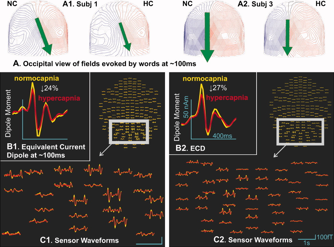
This paper explores how changes in the brain's hemodynamic and metabolic environment influence underlying neural activity. Using magnetoencephalography (MEG) during auditory pattern recognition and visual semantic tasks, the study demonstrates that mild hypercapnia induces a significant, widespread decrease in both early sensory and later cognitive event-related fields without altering behavioral performance. The findings suggest that this cortical suppression acts as a homeostatic mechanism to downregulate neuronal activity when blood flow cannot easily meet metabolic demands, which provides critical insights for refining the calibration of BOLD fMRI hemodynamic models.
Links
BibTeX tap to expand
@article{Thesen_hypercapniaBOLD_2012,
author = {Thesen, Thomas and Leontiev, Oleg and Song, Tao and Dehghani, Nima and Hagler Jr, Donald J and Huang, Mingxiong and Buxton, Richard and Halgren, Eric},
title = {Depression of cortical activity in humans by mild hypercapnia},
journal = {Human Brain Mapping},
volume = {33},
number = {3},
pages = {715-726},
keywords = {cerebral blood flow, cerebral metabolic rate for oxygen, blood oxygenation level-dependent response, functional magnetic resonance imaging, magnetoencephalography, temporal lobe, prefrontal cortex, occipital lobe, carbon dioxide, arterial spin labeling},
doi = {https://doi.org/10.1002/hbm.21242},
url = {https://onlinelibrary.wiley.com/doi/abs/10.1002/hbm.21242},
eprint = {https://onlinelibrary.wiley.com/doi/pdf/10.1002/hbm.21242},
year = {2012}
}
Code & Data
The room
Abstract
The effects of neural activity on cerebral hemodynamics underlie human brain imaging with functional magnetic resonance imaging and positron emission tomography. However, the threshold and characteristics of the converse effects, wherein the cerebral hemodynamic and metabolic milieu influence neural activity, remain unclear. We tested whether mild hypercapnia (5% CO2) decreases the magnetoencephalogram response to auditory pattern recognition and visual semantic tasks. Hypercapnia induced statistically significant decreases in event-related fields without affecting behavioral performance. Decreases were observed in early sensory components in both auditory and visual modalities as well as later cognitive components related to memory and language. Effects were distributed across cortical regions. Decreases were comparable in evoked versus spontaneous spectral power. Hypercapnia is commonly used with hemodynamic models to calibrate the blood oxygenation level-dependent response. Modifying model assumptions to incorporate the current findings produce a modest but measurable decrease in the estimated cerebral metabolic rate for oxygen change with activation. Because under normal conditions, low cerebral pH would arise when bloodflow is unable to keep pace with neuronal activity, the cortical depression observed here may reflect a homeostatic mechanism by which neuronal activity is adjusted to a level that can be sustained by available bloodflow. Animal studies suggest that these effects may be mediated by pH-modulating presynaptic adenosine receptors. Although the data is not clear, comparable changes in cortical pH to those induced here may occur during sleep apnea, sleep, and exercise. If so, these results suggest that such activities may in turn have generalized depressive effects on cortical activity.
Citing
If you use this code or build on these ideas, please cite the paper using the BibTeX entry above.
Doors · concepts in this room
Related rooms
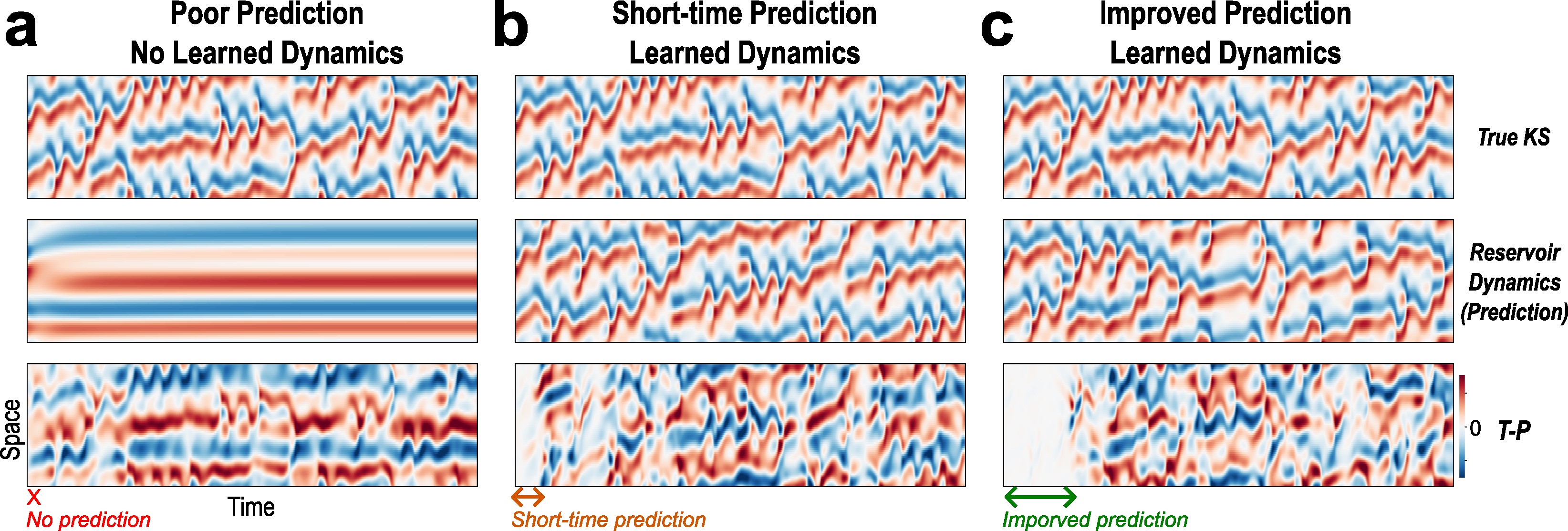
Evolutionary Optimization Reveals Structural Constraints on Reservoir Architecture for Spatiotemporal Chaos
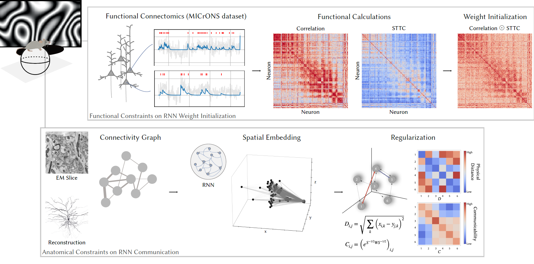
Harnessing cortical geometry, wiring, and function as inductive biases for recurrent neural networks